The Virtual Microscope is a NASA-funded project that provides simulated scientific instrumentation for students and researchers worldwide as part of NASA's Virtual Laboratory initiative. This site serves as home base for the Imaging Technology Group's contributions to that project—namely virtual microscopes and the multi-dimensional, high-resolution image datasets they view. Currently we provide 90 samples totaling over 62 gigapixels of image data. The Virtual Microscope, which is available for free download supports functionality from electron, light, and scanning probe microscopes, datasets for these instruments, training materials to learn more about microscopy, and other related tools.
Our Virtual Instruments
Our virtual instrument code currently supports data from three different instruments in our Microscopy Suite: a Philips Environmental Scanning Electron Microscope (ESEM), a Fluorescence Light Microscope, and an Atomic Force Microscope. We have also adapted a high-resolution Digital SLR with a 5x magnifying macro lens to capture some specimens, as well as included some artistic renderings of microscopy data.
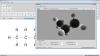
The virtual microscope aims to present the user with a method for exploring these pre-captured image data as if they were using the real instrument in real-time. To fulfill this goal, the virtual microscope provides the ability to load/unload specimens, to navigate to any point on that specimen, to change magnification, to adjust image parameters (contrast and brightness), to change focus, to analyze elemental composition, to measure features, and to render data in three dimensions. Additionally, the interface allows experts and laypeople alike to annotate specimens and/or load previously-created annotations.
Beyond the user interface, we have written a backend suite of custom software for the various tasks involved in collecting and processing the image data. This includes automated data collection of the thousands of images it takes to describe a single specimen, and routines for stitching and blending those tiled image datasets.
Microscope Training
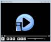
SEM Manual ScrenshotAs part of our educational mission, we have produced animations that teach the basics of electron, light, and scanning probe microscopy, videos detailing sample preparation for those instruments, videos of interviews with graduate students about their career paths in the sciences, and help videos about how to use our application. These materials animations use a multitude of media to explore various topics relevant to the theory and craft behind the images.
The Virtual Microscope
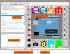
The Virtual Microscope is a Java application that supports interactive viewing of high-resolution, multi-dimensional image datasets from various microscopes. We currently support data from a Philips Environmental Scanning Electron Microscope (ESEM), and a Fluorescence Light Microscope.
Interface Controls
The interface provides a simulation of our actual microscope interface(s). Controls common to all datasets include brightness, magnification (usually up to 1800x), navigation, measurement, and annotation. SEM samples include a contrast control, and some also include a focus control.
The navigation and magnification controls allow the user to explore any point of interest on the sample. This differs from other virtual microscopes that have been produced which provide only a few 'hot spots' that can be magnified and explored.
Focus
The focus control is a real focal plane adjustment instead of a blurring of the image. Our automated data capture software (see below) allows us to capture an arbitrary number of focal planes. Thus, the focus control on the Virtual Microscope allows the user to glide through those planes interactively, bringing items closer and farther into focus. This is particularly useful with thick specimens viewed in the SEM.
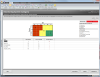
Annotations
We have included a full set of specimen annotation tools so that experts and laypeople alike can mark up the datasets for future reference. These annotations are useful for teaching details about the data, as well as for allowing teachers to ask students to identify data features as homework. The annotations can be saved and reloaded as XML files.
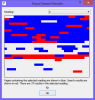
Automated Data Collection
It can take thousands of images to fully describe a single specimen so that the user can explore any point at varying magnifications with multiple focal planes. As a result, some of our datasets are many gigabytes (GB) in size. In order to accurately and sanely collect this data, we have written various software programs that automatically control the instruments.